
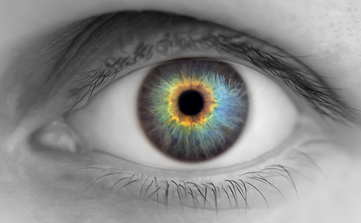
I would like to welcome you to the summer edition of European Ophthalmic Review, which features timely reviews and updates in glaucoma and surgical techniques, ocular surface, dry eye disease, diabetic macula oedema, optic neuritis and uveal melamona. My featured article entitled ‘Ocular Surface Disorders and Cataract and Refractive Surgery Success’ highlights the importance of the study of the meibomian glands in patients undergoing cataract or refractive surgery. In recent years we have seen a significant number of patients complain of poor results even when surgery was successful. They usually have poor visual quality and subjective complaints, with a low degree of satisfaction. In many of these cases, ocular surface disorders were not diagnosed before surgery, which are the cause of poor results. One of the problems we found when studying the ocular surface is that we do not always have the appropriate criteria and the most effective methods to detect a possible disorder. Usually we just study the tear break-up time, tear secretion and vital dyes of the cornea and conjunctiva, but we sometimes forget to assess the structure of the tear, the lipid layer and the state of the meibomian glands. Today we have new technologies that allow us to analyse these factors, especially the ability to view the meibomian glands, their number and appearance, as well as the lipid layer, the tear film distribution, even in a dynamic way, which provides more information to the assessment of their participation in the potential damage to the ocular surface. In our experience we have seen that the possibility of studying the ocular surface and the state of the meibomian glands is critical to prepare patients before cataract or refractive surgery. Improved visual results and the degree of satisfaction has increased significantly, so we recommend incorporating this practice into the patients’ protocol for anterior segment surgeons. European Ophthalmic Review would like to take this opportunity to thank all participants on this edition, from organisations to individuals. A special thanks goes to our editorial board for their continuing support and invaluable guidance and the biggest thanks are reserved for the expert authors, who spared precious time and effort to produce a perceptive selection of articles. This expert discussion and the wide variety of topics covered ensure there is much of interest for every reader and we hope you find this edition as useful and insightful as those before it.
Foreword – European Ophthalmic Review, 2013;7(1):5
Article
Further Resources

Trending Topic
On 28 May 2024, enrolment in phase III clinical trials for sozinibercept in neovascular age-related macular degeneration (nAMD) was completed.1 These trials include two large multicentre, double-masked, randomized controlled trials (RCTs): COAST (OPT-302 with aflibercept in neovascular age-related macular degeneration; ClinicalTrials.gov identifier: NCT04757636) and ShORe (OPT-302 with ranibizumab in neovascular age-related macular degeneration; ClinicalTrials.gov identifier: NCT04757610).2,3 These trials represent one of the largest phase […]
On 28 May 2024, enrolment in phase III clinical trials for sozinibercept in neovascular age-related macular degeneration (nAMD) was completed.1 These trials include two large multicentre, double-masked, randomized controlled trials (RCTs): COAST (OPT-302 with aflibercept in neovascular age-related macular degeneration; ClinicalTrials.gov ...

We are pleased to present the latest edition of touchREVIEWS in Ophthalmology. In this issue, we offer a series of engaging editorials, in-depth review articles and insightful original research highlighting some of the latest breakthroughs, innovations and practical insights in ...

Age-related macular degeneration (AMD) is a chronic deterioration and dysfunction of the outer retinal tissue and Bruch’s membrane (BrM). It is the leading cause of vision loss in people older than 60 years and is estimated to affect 288 million people ...

Neovascular age-related macular degeneration (nAMD) and diabetic macular oedema (DME) are two leading causes of visual impairment and blindness in the USA.1,2 Faricimab is the first bispecific antibody approved by the US Food and Drug Administration in ophthalmology and was ...
Age-related macular degeneration (AMD) is one of the main causes of irreversible vision loss in ageing populations worldwide.1 In 2019, the US recorded 1.49 million people aged 40 years or older with late-stage AMD, reflecting a crude prevalence rate of 0.94%.2 Geographic atrophy (GA), ...

Age-related macular degeneration (AMD) is a progressive eye condition that affects millions of people worldwide and poses a significant threat to vision in individuals over the age of 50.1 Of the two types of AMD, wet AMD, which is characterized by ...

With the introduction of monoclonal antibody inhibitors for all isoforms of vascular endothelial growth factor (VEGF)-A, which were first presented in September 2005 with off-label use of bevacizumab, significant anatomical and visual improvements for neovascular age-related macular degeneration (nAMD) became ...

The subspecialty of the retina has been transformed by anti-vascular endothelial growth factor (anti-VEGF) medications over the last two decades.1 These include bevacizumab (Avastin®, Genetech, San Francisco, CA, USA), ranibizumab (Lucentis®, Genentech, San Francisco, CA, USA), aflibercept (Eylea®, Regeneron Pharmaceuticals, ...

Age-related macular degeneration (AMD) is a leading cause of vision impairment and legal blindness in developed countries, with a projected estimate of 288 million cases in 2040.1 AMD can be classified as exudative or non-exudative. The early AMD-related lesion is represented by ...

Age-related macular degeneration (AMD) is characterized by age-associated thinning of the macula and formation of drusen. It has substantial global prevalence, especially in the older population, and it is the primary cause of permanent loss of vision in individuals 50 years ...
The development of anti-vascular endothelial growth factor (VEGF) agents as intravitreal treatments for neovascular age-related macular degeneration (nAMD) has drastically improved the vision outcomes for this disorder. Ranibizumab, a recombinant, humanized monoclonal antibody that targets VEGF-A, was approved in 2006 by ...
Neovascular age-related macular degeneration (nAMD) is characterized by the growth of abnormal blood vessels extending through Bruchs membrane in the macular region.1 This macular neovascularization (MNV) can be classified based on localization as either type 1, type 2, type 3 or mixed lesions.2 ...
Log into your Touch Account
Keep track of your clinical interests and newsletter subscriptions.
Sign up with an Email
Or use a .
Register now for FREE access
Already registered? Login below.
