Ophthalmologists, like all medical professionals, strive to base their practice on solid evidence. However, even in our field, certain beliefs have taken root more through tradition and repetition than through rigorous scientific scrutiny. Margolis and Galor published their editorial in which they debunked six myths related to the anterior segment.1 Their work motivated our critical thinking, and we bring to light 10 additional myths that continue to shape everyday clinical decisions. It is worth emphasizing that much of the evidence challenging these myths has emerged only recently – in many cases, within the past 18–24 months – and it is therefore unsurprising that established practice patterns have yet to reflect these findings. By examining why these myths endure despite evidence to the contrary, we hope to encourage a shift toward practices more firmly grounded in current research. Addressing these misconceptions is crucial not only for improving patient outcomes but also for understanding the gaps in medical education and the dissemination of knowledge.
Myth 1: mannitol is ineffective in vitrectomized eyes
Many of us learned early in our training that mannitol’s pressure-lowering effect relies solely on shrinking the vitreous cavity. This seemingly logical explanation led to the widespread teaching that mannitol would be ineffective in vitrectomized eyes. However, recent evidence challenges this long-held belief. Serhan et al. conducted a systematic review and meta-analysis of 5 studies, including 236 eyes (145 vitrectomized and 91 non-vitrectomized), to evaluate the efficacy of intravenous mannitol in reducing intraocular pressure (IOP).2 Their findings revealed significant IOP reductions in both vitrectomized and non-vitrectomized eyes across multiple time points. In vitrectomized eyes, IOP decreased by 19% at 30 minutes (min), 16.7% at 60 min, 24.3% at 90 min, 27.4% at 2 hours (h), 30.8% at 3 h and 30% at 4 h. These results demonstrate that mannitol’s efficacy is not dependent on the presence of vitreous, challenging the notion that it is ineffective in vitrectomized eyes. The study highlights the importance of reconsidering mannitol as a viable option for managing acute IOP elevation in post-vitrectomy patients, ensuring optimal perioperative care.
Myth 2: cyclopentolate is essential for pediatric cycloplegic refraction
The preference for cyclopentolate in pediatric cycloplegic refraction has become nearly standard practice in our field, often justified by its stronger cycloplegic effect. However, recent evidence suggests that tropicamide, a shorter acting agent, may achieve comparable refractive results in select patient populations while offering practical advantages. A randomized clinical trial involving 185 eyes from 94 children aged 3–16 years compared the efficacy of cyclopentolate 1% and tropicamide 1% specifically in pediatric patients with brown irides.3 The study found that the mean spherical equivalent (SE) after cycloplegia was 1.07 ± 5.2 diopters for cyclopentolate and 0.96 ± 5.1 diopters for tropicamide, with no clinically significant difference between the two agents (p<0.001). The mean change in SE (ΔSE) after cycloplegia was 1.15 ± 1.2 for cyclopentolate and 1.04 ± 1.2 for tropicamide, with a statistically insignificant difference of 0.11 ± 1.2 (p<0.001). Notably, the greatest effect of both agents was observed in hyperopic eyes, with ΔSE of 1.54 ± 1.4 for cyclopentolate and 1.39 ± 1.4 for tropicamide.
These findings suggest that tropicamide may be an effective and safe alternative to cyclopentolate for cycloplegic refraction in pediatric patients without strabismus and with brown irides, regardless of their refractive error status – as the study itself acknowledges. Generalization of these results to children with lighter irides or to patients with strabismus should be made with caution, as the study’s authors note that patients with strabismus were specifically excluded. In the population without strabismus and with brown irides studied, the shorter duration of action and better tolerability of tropicamide represent meaningful advantages, reducing the burden of prolonged pupillary dilation and photophobia for both patients and families.
Myth 3: aspirin must be stopped before cataract surgery
The practice of discontinuing aspirin before cataract surgery exemplifies how theoretical risks can overshadow practical evidence. While concern about hemorrhagic complications is understandable, large-scale studies show that continuing aspirin does not significantly increase the risk of sight-threatening bleeding events. A systematic review and meta-analysis of 65,196 patients found that aspirin continuation only raised the risk of subconjunctival hemorrhage (risk ratio: 1.74; 95% confidence interval [CI]: 1.22–2.50; p=0.002), with no significant increase in serious complications such as hyphema, retrobulbar hemorrhage or vitreous hemorrhage.4 Meanwhile, the thrombotic risks of stopping aspirin are real, particularly for patients with cardiovascular conditions.
This myth has far-reaching implications given the volume of cataract surgeries performed globally and the increasing prevalence of antiplatelet therapy in our aging population. The decision to stop aspirin often triggers a cascade of consultations with primary care physicians and cardiologists, potentially delaying surgery. More concerning is the risk of thrombotic events in patients who stop their antiplatelet therapy, especially those with recent coronary stents or other high-risk conditions. Yet, many clinicians still routinely ask patients to stop aspirin, creating unnecessary anxiety and risk. This gap between evidence and practice raises important questions about how we evaluate and balance competing risks in clinical decision-making. The evidence supports continuing aspirin perioperatively, ensuring patient safety while minimizing unnecessary disruptions to their care.
Myth 4: prostaglandin analogs increase cystoid macular edema risk
Despite being our first-line agents for glaucoma, prostaglandin analogs (PGAs) continue to carry the stigma of potentially causing cystoid macular edema (CME). This concern has led many clinicians to avoid these medications in patients with risk factors for CME, even though current evidence fails to support this association. A large database study of 39,948 patients newly prescribed glaucoma medications compared the incidence of CME among users of PGAs, beta-blockers (BBs), alpha agonists (AAs) and carbonic anhydrase inhibitors (CAIs).5 The results showed that the incidence of CME was lowest among PGA users (0.13%) compared with BBs (0.65%), AAs (0.55%) and CAIs (1.76%). After adjusting for sociodemographic factors, users of BBs, AAs and CAIs had significantly higher odds of developing CME compared with PGA users (p<0.001 for all comparisons).
This pattern is further corroborated by multiple independent analyses. A systematic review and meta-analysis by Özyol et al., which analyzed nine studies of perioperative PGA use in cataract surgery, found that continuing PGAs perioperatively did not significantly increase CME risk compared with discontinuing them (OR=1.32; 95% CI: 0.49–3.51; p=0.582), and that PGA use did not significantly increase CME risk compared with other antiglaucomatous medications (odds ratio [OR]=2.29; 95% CI: 0.84–6.23; p=0.103).6 The authors concluded that discontinuing PGAs perioperatively in eyes without known CME risk factors has no clinically significant effect on reducing postoperative CME. Similarly, a systematic review of randomized controlled trials by Santamaria et al., which included four randomized controlled trials, found no causal relationship between PGA use and pseudophakic CME following uneventful cataract surgery, concluding that PGAs do not need to be suspended in patients without known risk factors.7
The persistence of this concern highlights how case reports and theoretical mechanisms can sometimes carry more weight in daily practice than larger, more rigorous studies. The impact extends beyond individual patient care to influence clinical trial design and drug development. Many studies still exclude patients on PGAs from CME-risk populations, potentially limiting our understanding of these medications’ true safety profile. These convergent large-scale findings should provide clinicians with greater confidence in prescribing PGAs as safe and effective first-line treatments for glaucoma, without reflexive concern about CME risk in the majority of patients.
Myth 5: prostaglandin analogs exacerbate uveitis in patients with pre-existing inflammation
A nuanced but widely held clinical belief – distinct from the concern about de novo CME – is that PGAs may exacerbate intraocular inflammation and make uveitis harder to control in patients with a history of uveitic disease. This theoretical risk, rooted in the understanding that prostaglandins are metabolites of arachidonic acid and therefore assumed to be pro-inflammatory, has led many clinicians to withhold PGAs from patients with uveitic glaucoma, precisely the population most in need of effective IOP lowering. However, evidence from large patient databases and targeted clinical studies tells a different story.
A retrospective study of 67,517 patients with glaucoma newly started on topical glaucoma therapy evaluated the incidence of uveitis among users of PGAs, BBs, AAs and CAIs.8 The overall incidence of uveitis was lowest among PGA users (0.32%) compared with BBs (1.95%), AAs (1.63%) and CAIs (1.68%). After adjusting for sociodemographic factors, users of BBs, AAs and CAIs had significantly higher odds of developing uveitis compared with PGA users (p<0.001 for all comparisons). Critically, in a secondary analysis of patients with a pre-existing history of chronic uveitis, these trends persisted – patients prescribed PGAs experienced lower rates of uveitic flares than those receiving other classes of glaucoma medications.
The mechanistic basis for PGA safety in uveitic eyes is also becoming clearer. In uveitis, the trabecular pathway is significantly compromised due to inflammation, pigment dispersion and the effects of corticosteroid use. PGAs, which act primarily through the uveoscleral outflow pathway, are therefore theoretically well suited for lowering IOP in this context.8 A pilot study by Taylor et al. found that PGAs do not significantly alter the expression of conjunctival inflammatory markers in patients with uveitic glaucoma, providing a cellular and molecular basis for the observed clinical safety.9 These findings, taken together, challenge the widely held concern that PGAs are contraindicated or harmful in patients with established uveitis and support their consideration as an effective IOP-lowering option in this challenging population – with appropriate individual clinical judgment and monitoring.
Myth 6: strict face-down positioning is essential after macular hole surgery
Few postoperative instructions are as burdensome to patients as face-down positioning after macular hole surgery. We have long insisted upon it based on theoretical benefits. However, evidence from both recent studies and long-term clinical experience challenges this practice. A Cochrane review of 8 randomized controlled trials involving 709 eyes found no significant difference in macular hole closure rates between patients who maintained face-down positioning and those who did not.10 Among patients with larger macular holes (≥400 μm), the closure rate was 94% in the face-down group compared with 84% in the non-face-down group. For smaller holes (<400 μm), the closure rate was 100% in the face-down group versus 96% in the non-face-down group. Similarly, Paul E. Tornambe, MD, in his 15-year review of macular hole repair without face-down positioning, reported a single-operation success rate of 92% in the no-face-down group compared with 90% in the face-down group. Tornambe’s findings emphasize that the buoyancy of the gas bubble, rather than strict positioning, is key to isolating the macular hole and promoting healing.11 He noted that patients who avoided face-down positioning experienced similar anatomical success rates, with the added benefit of reduced discomfort and improved quality of life.
The impact of this practice extends beyond patient discomfort to affect surgical decision-making and resource allocation. Many elderly or physically limited patients may be denied surgery based on concerns about positioning compliance, while others may require extended stays in skilled nursing facilities solely to ensure adherence. The evidence suggests that modified positioning regimens can achieve equivalent results, reducing the burden on patients and healthcare systems alike. These findings support a reappraisal of strict face-down positioning in favor of more individualized, patient-centered postoperative protocols.
Myth 7: superior laser peripheral iridotomy location prevents dysphotopsia
The practice of placing laser peripheral iridotomy (LPI) at the 12 o’clock position to prevent dysphotopsia illustrates how anatomical reasoning can lead practice astray. While it seems logical that hiding the iridotomy behind the upper lid would reduce visual symptoms, systematic study of patient outcomes does not support this assumption. A systematic review of 1,756 eyes from 878 patients compared the incidence of dysphotopsia based on LPI location (superior, inferior or nasal/temporal).12 The analysis found no significant difference in the incidence of dysphotopsia among the location groups. Overall, the incidence of linear dysphotopsia was 2–3% regardless of LPI placement. Interestingly, the review also noted that LPI could resolve pre-existing halos and glare, suggesting that the procedure may have some beneficial effects on visual symptoms.
Beyond this, randomized trial data specifically comparing temporal and superior placement favor temporal locations for outcomes. Vera et al. conducted a prospective, randomized, paired-eye trial in which 208 patients received temporal LPI in one eye and superior LPI in the other.13 New-onset linear dysphotopsia was significantly less common in the temporal group (2.4%) than in the superior group (10.7%; p=0.002). Notably, 6.5% of eyes with superior LPI reported linear dysphotopsia despite complete eyelid coverage of the iridotomy, underscoring that lid coverage alone is an insufficient protective mechanism. These findings are further supported by a large retrospective study of 2,385 eyes by Singh et al., which found higher total dysphotopsia rates with superior LPI (11.2%) compared with temporal LPI (8.0%).14
Moving beyond the assumption of superior placement allows for more individualized approaches to iridotomy placement based on each patient’s lid position, iris architecture and anterior chamber depth. The accumulating evidence supports temporal placement as a preferred location in appropriate candidates, offering both equivalent therapeutic efficacy and a more favorable visual symptom profile.
Myth 8: symptomatic floaters should be treated by drinking liters of water and oral supplementation
The management of vitreous floaters remains a topic of on-going research and debate in ophthalmology. Although increased water intake (2–3 L daily) and oral supplementation with sodium, potassium and collagen are not taught in formal medical curricula, these recommendations have gained significant traction among patients through social media, online health forums and anecdotal testimonials. Despite their popularity in patient communities, these claims lack scientific validation. While proper hydration contributes to overall health, no clinical evidence supports its direct role in floater resolution.
The only non-surgical, dietary treatment for vitreous floaters with published clinical data comes from Ma et al., who conducted a clinical trial evaluating mixed fruit enzymes.15 Their study demonstrated promising results, with floater disappearance rates of 55%, 62.5% and 70% for daily doses of 1, 2 and 3 capsules, respectively (p<0.001). In cases of vitreous hemorrhage-induced floaters, disappearance rates were 18%, 25% and 56% (p<0.001) with increasing doses.
Currently, the only scientifically backed methods for treatment of vitreous floaters are pars plana vitrectomy (PPV) and neodymium-doped yttrium aluminium garnet (Nd:YAG) laser vitreolysis.16 While PPV demonstrates high patient satisfaction rates (85–100%), it carries potential risks such as retinal detachment and cataract formation. Nd:YAG laser vitreolysis offers a less-invasive alternative, though with more modest success rates of 35.8% compared with PPV’s 93.3%.16 The growing prevalence of unvalidated floater ‘treatments’ in patient-facing media underscores the need for ophthalmologists to proactively counsel patients about evidence-based options during clinical encounters.
Myth 9: myopia progresses significantly during pregnancy
A common concern leads some women to delay refractive surgery out of fear that myopia will worsen during pregnancy, thereby invalidating surgical results. Ong et al. conducted a comprehensive longitudinal analysis of 269 women over 8 years, finding no significant associations between pregnancy and myopia progression.17 While pregnancy was associated with a slight increase in lens thickening (0.004 mm/year for each full-term pregnancy; 95% CI: 0.002–0.007), this did not translate to significant refractive changes. The study comprehensively analyzed multiple ocular parameters – including SE, axial length, corneal radius and lens thickness – finding no significant pregnancy-related changes except for lens thickness.
These findings specifically challenge the assumption that pregnancy induces clinically meaningful myopic progression. However, it is important to acknowledge the boundaries of this evidence: the study by Ong et al. addresses refractive stability during pregnancy and does not directly examine refractive surgery outcomes, corneal biomechanical changes associated with pregnancy or healing and recovery in the postoperative period.17 Decisions about the timing of refractive surgery remain multifactorial and should be individualized. Within the specific domain of myopia progression, however, the available longitudinal data do not support significant refractive deterioration during pregnancy, and this particular concern need not be the sole reason to indefinitely defer surgery in appropriate candidates.
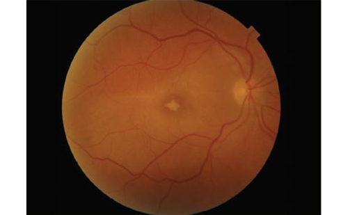
Myth 10: symptomatic floaters always indicate a diagnosis of posterior vitreous detachment
The routine attribution of floaters to posterior vitreous detachment (PVD) in clinical practice reflects an important diagnostic oversimplification that may be better described as a pattern of overdiagnosis rather than mismanagement. Ripa et al. analyzed 1,472 eyes using widefield optical coherence tomography (OCT) and found that PVD was present in only 66% of patients presenting with floaters.18 Specifically, they detected complete PVD in 571 eyes (39%), partial PVD in 69 eyes (5%) and no PVD in 560 eyes (38%). Standard macular OCT could raise suspicion for complete PVD in only 14% of cases, highlighting the limitations of conventional imaging in this context.
The practical implication is that a substantial proportion of patients presenting with floaters – roughly one in three in this cohort – have no detectable PVD on widefield OCT. Relying on symptoms alone to presume a PVD diagnosis risks misclassifying cases where the floaters arise from other vitreous pathology, including vitreous syneresis, asteroid hyalosis or hemorrhage. The study also found that symptom severity correlates more strongly with opacity size, density and location than with the mere presence or absence of PVD, further reinforcing the need for thorough imaging-based evaluation rather than a presumptive diagnosis. Widefield OCT enables precise characterization of the vitreoretinal interface, including the distinction between complete and partial PVD, and should be incorporated into the workup of symptomatic patients where clinical uncertainty exists.
Discussion
The myths reviewed in this report share a common thread: they arose from plausible biological reasoning, case-level observations or traditional teaching and persisted not because of evidence in their favor, but because the evidence against them is relatively recent and has not yet been widely disseminated or integrated into clinical practice. Indeed, a defining feature of many of the studies cited here is their novelty – much of the contradictory evidence was published within the past 18–24 months. This temporal context is essential for understanding why these myths remain influential: it is not that the evidence has been ignored, but that the systems for translating new findings into updated practice have inherent lag.
This reality places a dual responsibility on the field. Educators must teach residents not only what to do but how to evaluate evidence critically – to ask what assumption underlies a teaching, what the quality of the supporting data is and whether that data have been tested against larger or more rigorous investigations. Practitioners, in turn, must remain open to revising well-established habits when new evidence warrants it. This is rarely straightforward: entrenched practices carry institutional momentum, and changing them requires more than the publication of a single study.
Some of the myths reviewed here carry clearer and more robust evidence bases than others. The data on mannitol in vitrectomized eyes, PGA safety with respect to CME and uveitis and aspirin continuation before cataract surgery now come from large-scale database studies and systematic reviews that substantially shift the burden of proof. Other areas – such as the optimal approach to cycloplegic refraction in diverse pediatric populations or floater management – require further study before broad practice changes are warranted. Acknowledging this variability in evidence quality is itself part of rigorous critical appraisal.
Effective knowledge translation requires more than awareness. It requires the integration of new findings into clinical guidelines and decision-support tools, the systematic review of practice patterns as part of quality improvement efforts and educational programs that cultivate a culture of questioning assumptions. By highlighting these examples, we aim to encourage colleagues to apply the same scrutiny to other aspects of their practice. Our field progresses not only through new discoveries but also through the willingness to reassess practices that may be rooted more firmly in tradition than in current evidence, regardless of how long those practices have been observed.