To view the full article in PDF or eBook formats, please click on the icons above.
There has been increasing interest in improving imaging technologies to identify and grade early age-related maculopathy (ARM).
Recently, advances in optical coherence tomography (OCT) has allowed a virtual biopsy of the retina and identification of different types of drusen in relation to early ARM. Several investigators have improved our understanding of both the qualitative and quantitative analyses of drusen, the clinical hallmark of ARM. The purpose of this article is to critically review the current literature inOCT in the evaluation of early ARM. Although seemingly useful and ideal, the current analysis of the retina using OCT is highly complex and lacking. It is currently too premature to use OCT as a grading tool for early ARM. However, the authors are hopeful that the rapid progress in this field will enable the use of an OCT-defined outcome measure in early ARM clinical trials in the foreseeable future.
To view the full article in PDF or eBook formats, please click on the icons above.
Trending Topic
Welcome to the latest edition of touchREVIEWS in Ophthalmology, and my first as the journal’s Editor-in-Chief. In this issue, we are delighted to present a series of compelling articles providing insights into some of the cutting-edge developments in this diverse and ever-evolving field. Dhanashree Ratra and Aashna Ratra open the edition with the first in […]
Oculoplastic surgeons are often faced with the diagnostic dilemma of abnormal tissues within the orbit and adnexal structures. In many cases, the associated signs, symptoms and radiological appearances allow a tentative differential diagnosis to be formulated. However, most often, diagnostic ...
Optical coherence tomography (OCT) is a revolutionary tool that plays a key role in the management of various retinal pathologies. The salient features of OCT are its non-invasive nature and its ability to image intraocular structures in vivo with resolution ...
The optic nerve head (ONH) is the region where retinal ganglion cells (RGCs) leave the eyeball via the scleral canal. It also consists of blood vessels, glial, and connective tissue. Glaucoma is a progressive optic neuropathy characterized by the loss ...
Q: What is the optimal procedure for pre- and intraoperative imaging of the eye during toric IOL implantation? Toric IOLs have perhaps enjoyed the greatest reproducible success of any premium lens type in use over the past decade. However, their ...
Spectral domain optical coherence tomography (SD-OCT) is a very useful non-invasive imaging method which is used in the diagnosis and follow-up of various retinal and choroidal diseases involving macular region.1–3 In 2014, at International Nomenclature OCT (INOCT) Panel, Staurenghi et al.4 ...
Many important eye diseases manifest themselves in the retina, one of the most prevalent of these is diabetic macular oedema. Retinal imaging and image analysis have developed rapidly over the past 10 years, and image analysis is starting to play an ...
Diabetic retinopathy (DR) is one of the leading causes of adult vision loss in the developed world.1,2 Epidemiological and demographic factors, including the rising rates of diabetes related to obesity and an aging population, are driving the incidence of diabetic ...
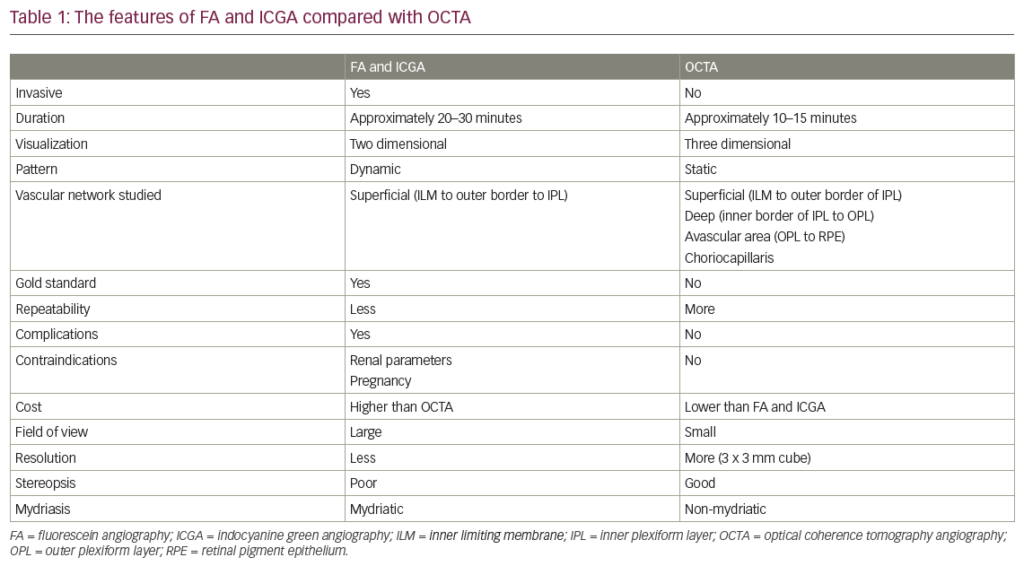
Imaging techniques in the diagnostic field of the eye have undergone a tremendous development over the last few decades. Fluorescein angiography (FA) as one of them, has been a key tool in the diagnosis of retinal diseases such as diabetic ...
Optical coherence tomography angiography (OCTA) is a type of noninvasive angiography without a contrast agent, which provides detailed assessment of the retinal and choroidal vasculature by detecting motions of erythrocytes and visualising blood flow using serial optical coherence tomography (OCT) ...
Imaging techniques in the diagnostic field of the eye have undergone a tremendous development over the last few decades. Fluorescein angiography (FA) as one of them, has been a key tool in the diagnosis of retinal diseases such as diabetic ...
Optical coherence tomography angiography (OCTA) is a relatively new diagnostic technique that is able to provide a rapid, dyeless and noninvasive three-dimensional reconstruction of perfused vessels of retina and choroid. The fundamental concept of OCTA is that in a static ...
In 2015, a novel imaging technology called optical coherence tomography angiography (OCTA) became available for clinical use. The features of many disorders, including diabetic retinopathy (DR), choroidal neovascularization (CNV), macular telangiectasia type 2, retinal vascular occlusion, and sickle cell retinopathy have been ...
Explore the latest in medical education and stay current in your field. Create a free account to track your learning.