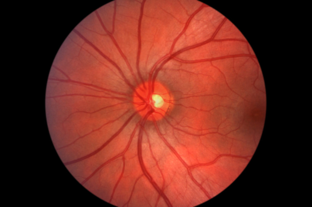
Diabetic retinopathy remains the major cause of blindness in working-age adults in developed nations. Diabetic retinal lesions are still reversible at the initial stage of mild non-proliferative diabetic retinopathy, opening real opportunities for effective intervention. Four main alterations characterise the early stages of diabetic retinopathy:
• microaneurysms/haemorrhages;
• alteration of the blood–retinal barrier (BRB);
• capillary closure; and
• alterations in the neuronal and glial cells of the retina.
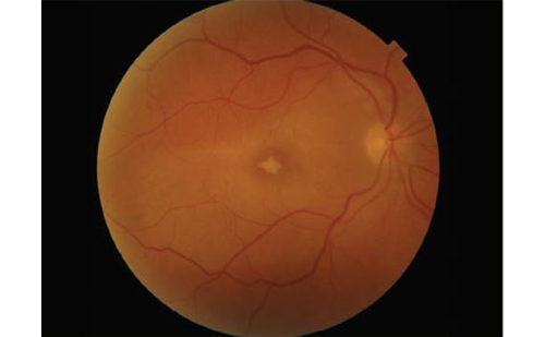
These alterations may be monitored by red-dot counting on eye fundus images, and by leakage and retinal thickness measurements.1,2 A combination of these methods through multimodal macula mapping has contributed to the identification of three phenotypes2 showing different patterns of evolution:
• pattern A, including eyes with reversible and relatively little abnormal fluorescein leakage, a slow rate of microaneurysm formation and normal foveal avascular zones;
• pattern B, including eyes with persistently high leakage values and high rates of microaneurysm accumulation; and
• pattern C, including eyes with variable leakage, high rates of microaneurysm accumulation and abnormal foveal avascular zones.