In a recently published study,1 we proposed an objective method of measuring the refractive power change after laser-assisted in situ keratomileusis (LASIK) surgery. In common refractive practice, the differences in manifest refraction are used to calculate the refractive changes after corneal refractive procedures such as LASIK or laser-assisted epithelial keratomileusis (LASEK). Measurement of refraction error using an autorefractor is less precise than that achieved following LASIK, and depends on the optical zone and the spherical aberration created.
In a recently published study,1 we proposed an objective method of measuring the refractive power change after laser-assisted in situ keratomileusis (LASIK) surgery. In common refractive practice, the differences in manifest refraction are used to calculate the refractive changes after corneal refractive procedures such as LASIK or laser-assisted epithelial keratomileusis (LASEK). Measurement of refraction error using an autorefractor is less precise than that achieved following LASIK, and depends on the optical zone and the spherical aberration created.
One problem of manifest refraction is that it is dependent on the person who is undergoing the procedure; this problem is more pronounced with young hyperopic patients as they are more difficult to accommodate and, therefore, the manifest refraction may not be a good indicator of predictability after hyperopic LASIK (H-LASIK) or surface ablation. Also worth considering is a clinical situation where the patient receives a successful laser treatment but after a couple of months suffers refractive problems: is this a hyperopia that was not properly treated, is it a regression or is it a presbyopia progression? Additionally, in this way actual under- or overcorrections can be differentiated from residual manifest refractions due to wrongly intended corrections. In these situations, measuring the cornea surface before and after corneal refractive procedures may help to objectively discern what is actually changed during the procedure. The data can also be used to compare different platforms and surgeons.
Materials and Methods
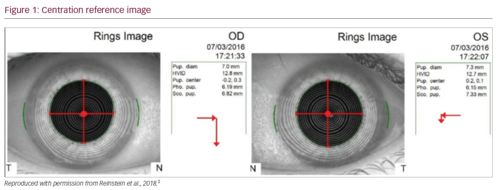
We designed a study in which we treated a consecutive group of hyperopia and hyperopic astigmatism with LASIK. The group consisted of 66 eyes, whose demographic data are shown in Table 1. All surgeries were performed by the same surgeon (DO). LASIK flaps were created with a superior hinge using a Carriazo-Pendular microkeratome2 (SCHWIND eye-tech-solutions GmbH, Kleinostheim, Germany). The ablation was carried out with an ESIRIS excimer laser3 (SCHWIND), and the ablation parameters were set for an optical zone of 6.25mm and a total zone of 7.75mm. We shifted the ablation centre to the corneal vertex centre4–8 using the pupillary offset (the distance between the pupil centre and the corneal normal vertex) measured by a videokeratoscope9 (Keratron Scout topographer, Optikon 2000 s.p.a., Italy). In the topography, the cornea vertex is always in the same place if the patient fixates correctly, and is reproducible and not dependent on the pupil diameter. The laser platform takes the centre of the pupil as the central target for ablation. If the ablation is shifted to the vertex, the pupil centre must be measured under the same photopic conditions as experienced during surgery, as the pupil centre changes if the pupil dilates, which means the vertex will not be calculated properly.
From the topography system, we take measurements of the flattest and steepest meridians of each zone at 3, 5 and 7mm, which are not necessarily 90° apart. The 3mm zone was examined using the simulated keratoscope reading (sim-K) and Maloney index analyses, showing that all corneas were normal. The resulting spherical equivalent (SE) was basically identical to that observed using the sim-K and Maloney indices. The progress of the spherical component was analysed at 5 and 7mm. We also calculated the optical errors represented by wavefront aberration at 6mm from the topography, described by Zernike polynomials and co-efficients in the Optical Society of America (OSA) standard.10,11
Results
Refractive Outcome
The pre-operative SE of this consecutive group of 66 eyes was +2.74 diopters (D) (range +0.75 to +4.75D) with a mean sphere of +3.07D (range +1.00 to +5.25D); the mean cylinder was -0.67D (range -4.00 to 0.00D). At three-month follow-up the mean SE was -0.09D (range -0.75 to +1.00D), the mean sphere was +0.02D (range -0.50 to +1.25D) and the mean cylinder was -0.21D (range -1.00 to 0.00D). The predictability was good, with 92% of the eyes within ±0.50D of the attempted correction; 100% were within ±1.0D. Referring to safety, no eye lost more than one line of best corrected visual acuity (BCVA) and one eye gained two lines of spectacle CVA after H-LASIK. From the data of a consecutive group of eyes we analysed the refractive change, which is defined as the vectorial difference in the astigmatism space of post-operative and pre-operative manifest refraction. The intended refraction and the intended refractive correction correlated (r2=0.91) and the slope of regression was 1.02, which is considered close to the ideal correction. Demographic data are represented in Table 1.
Corneal Topographical Changes
From the corneal topography, we analysed the different pre-operative and three month post-operative indices (see Tables 2 and 3). The different refractive indices used for the topography and the ablation planning – keratometric refractive index 1.3375 for the topographies, and corneal refractive index 1.376 for the ablations – were taken into account. Furthermore, the vertex distance used in ablation planning laser settings was considered.
The Maloney index correlated most significantly with the intended refractive correction (r2=0.81). The slope of the regression was 1.00, and therefore close to the ideal correction. The achieved sim-K change was significantly correlated with the intended refractive correction (r2=0.36) and the slope of regression was 0.92, and thus slightly undercorrected. The achieved power change at 5 and 7mm was not correlated with the intended refractive correction, meaning that the refractive change is at the centre of the cornea until 5mm. Furthermore, we found a slope regression of -0.17 at 7mm, indicating a myopic-like effect even for intended hyperopic corrections. We also calculated the asphericity changes12 and noted that treated corneas became more prolate after H-LASIK.
Discussion
In our consecutive group of eyes, H-LASIK with the Schwind ESIRIS platform showed good predictability, with 92% of the eyes having a post-operative refraction within ±0.50D of the attempted correction. The achieved refractive change was significantly correlated with the intended refractive correction (r2=0.91), and was close to the attempted correction.
By analysing the corneal topographical changes, a highly significant correlation between the resulting Maloney change and the intended refractive correction could be observed (r2=0.81), which was close to the ideal correction (see Figure 1). The sim-K was significantly correlated with the intended refractive correction (r2=0.36), and was still quite close to the ideal correction, and only slightly undercorrected.
Based on the good agreement between refractive and topographical changes, topographical indices could be used instead of manifest refraction to contrast the results of different surgeons or for nomogram optimisation. As topography is a highly reproducible method, in this way any undesired accommodative effects – such as different methods to determine the subjective refraction – can be directly avoided in the analysis and the influence of the technician or patient can be minimised. This is important, as the majority of hyperopic patients have the ability of a certain degree of accommodation, thus masking their hyperopia and disregarding the fact that presbyopia could be detected in the majority of our patient group, whose average age was 50 years. In cases of young patients manifesting hyperopia, refraction used to be understimated as an accommodative response helps these patients to achieve good visual acuities without full hyperopic correction. In this way, our topographical method of analysis allows surgeons to assess whether an under- or overcorrection is real or due to a wrong refraction or intended correction. We will also investigate in future studies whether the topographical data allow us to see whether there is a regression after a refractive procedure and whether this occurs at the centre or the periphery. Different ablation profiles will also be considered.
Based on our results, we suggest that the Maloney index is a better descriptor compared with the sim-K analysis because it showed lower scatter in our cases (r2=0.81 for Maloney versus r2=0.36 for sim-K). An explanation for this could be that the sim-K represents the flattest meridian when analysed at 3mm and takes the other principal meridian 90° away, independent of its curvature, so that it is not neccessarily the steepest meridian. Alternatively, we could take the steepest meridian at 3mm analysis and the meridian 90° away independently from its curvature, but again these are not neccessarily the flattest meridians. Another option is to consider the two meridians 90° away at 3mm analysis, which maximise the astigmatism and therefore are not neccessarily the flattest and steepest meridians. In ‘normal’ corneas, without irregular astigmatism, these three methods of analysis provide similar results. Sim-K analysis is a simple 2D cross-sectional analysis.Additionally, the Maloney index uses the inner 3mm zone to provide the best fit for the disc area to a spherocylindrical surface in 3D. Cylinder orientation defines the two principal meridians, and the sphere and cylinder provide the curvatures of the principal meridians. Other authors13 analysed the topographical changes after photorefractive keratectomy in myopia and also found a better correlation with the Maloney index compared with sim-K.
The achieved power changes at 5 and 7mm were not correlated with the intended refractive correction, and they showed a decreasing correction factor, indicating a progressive severe undercorrection, and at 7mm a myopic-like effect even for intended hyperopic corrections. The myopic-like behaviour can be explained by the fact that the treatments were planned at the 6.25mm optical zone, and our analysis addressed the 7mm zone, meaning that our analysis already included the transition zone area (TZ), where the hyperopic profiles showed a myopic-like shape. The good agreement between refractive and topographical changes at 3mm (with both Maloney and sim-K) can be explained by 3mm being a common pupil size during manifest refraction diagnosis. We induced negative sphericalaberration. Earlier studies on hyperopic treatments with excimer lasers also suggested an increase in negative spherical aberration.14–18 In our group of eyes, the induced corneal spherical aberration decreased on average by – 0.09μm per D for a 6mm pupil.
Spherical aberration is also directly related to multifocality, and confirms the topographical findings of multifocality. By analysing the slope of the cornea, we can also measure the multifocality. We observed a clear multifocality for both topographical and corneal wavefront analysis. Also, the central part of the cornea is slightly overcorrected (inducing a post-operative central myopia), and there is an extended depth of focus of about 1.25D (probably helping the patients to approach presbyopia). This may explain why in some cases the patients – although presbyopic – reported improved near vision after the procedure, confirmed by a reduced need for addition for reading (the majority of patients treated with hyperopic LASIK were presbyopic, so before surgery they required the addition of a positive sphere for reading; after surgery, they required less addition).
We used a classic treatment, where the induced negative spherical aberration and asphericity increases with the attempted correction. We expected less induction of aberrations in a profile that takes into account the loss of efficency at the periphery,19,20 and therefore such a profile will give us less multifocality. The knowledge of the resulting positive effect in presbyopic–hyperopic laser treatments may allow us to optimise the amount of spherical aberration that is induced. We were able to demonstrate that topographical changes of the Maloney index can effectively be used in analysing laser corneal refractive surgeries for hyperopia and avoiding undesired accommodative effects in the analysis. In this way, our topographical method of analysis is objective and allows surgeons to assess whether an under- or an overcorrection is real or due to a wrong refraction or intended correction. On these grounds, this method has the potential to be used as an objective method of analysis based on subjective manifest refractions for presentation of refractive surgery results or for optimisation of nomograms.
Based on the good agreement between refractive and topographical changes, topographical indices could be used instead of the manifest refraction to compare the results of different surgeons, or for nomogram optimisation.


