This website is intended for healthcare professionals only

Trending Topic

Macro shot of eye featuring holographic HUD graphics layered over pupil and cornea, neon light accents and sharp reflections, high tech cyber vision theme, ultra detailed 8k
7 mins

Trending Topic

Developed by Touch
Mark CompleteCompleted
BookmarkBookmarked

Corneal ectatic disorders, such as keratoconus, progressively weaken corneal integrity, leading to thinning, irregular astigmatism and visual deterioration.1 Typically progressive in nature, these ectasias result in increasingly thinner corneas, causing the cornea to protrude forward into a cone shape. This leads to increasing amounts of myopia and astigmatism – both regular and irregular – as the disease […]

Evaluation of the Optic Nerve and Retinal Nerve Fibre Layer in Myopic Individuals

Ahmad A Aref
< 1 min
Share
Facebook
X (formerly Twitter)
LinkedIn
Via Email
Mark CompleteCompleted
BookmarkBookmarked
Copy LinkLink Copied
Download as PDF
Published Online: Jan 23rd 2013 European Ophthalmic Review, 2012;6(5):280-282 DOI: http://doi.org/10.17925/EOR.2012.06.05.280
Select a Section…
1

Article

Abstract

Clinical discrimination between myopic tilted optic discs and glaucomatous optic neuropathy is often challenging, especially when

Abstract

Clinical discrimination between myopic tilted optic discs and glaucomatous optic neuropathy is often challenging, especially when
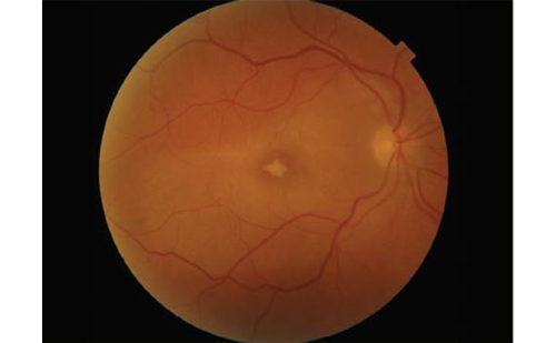
considering that myopia is a risk factor for the development of glaucoma. Myopic tilted discs are usually larger than average, with associated relative cupping and thinner neuroretinal rim tissue. Histopathological study has revealed thinner parapapillary retinal tissue in these eyes. Optical coherence tomography (OCT)-measured average retinal nerve fibre layer (RNFL) thickness has been found to decrease with longer axial length and higher myopic refractive error. Parapapillary RNFL quadrant and clock-hour analyses result in a higher false-positive rate in myopic eyes. Careful slit-lamp examination, quality baseline stereoscopic disc photographs and frequent serial visual field testing are essential to the follow-up of myopic individuals with suspected glaucoma. A novel diagnostic parameter, OCT-derived ganglion cell analysis, may prove to be useful in the diagnosis and follow-up of these individuals.

To view the full article in PDF or eBook formats, please click on the icons above.

2

Further Resources

Share
Facebook
X (formerly Twitter)
LinkedIn
Via Email
Mark CompleteCompleted
BookmarkBookmarked
Copy LinkLink Copied
Download as PDF
Close Popup