Corneal refractive surgery is a common choice for reducing the dependence of vision correction by glasses or contact lenses. However, as with any form of surgery or medical treatments, there is an inherent risk of complications. These risks, and their subsequent management, can vary widely depending on which technique is employed and the preoperative assessment.1,2 In order to mitigate, and even predict, the complications of surgery, it is essential to conduct a thorough screening for predisposing conditions.3 A comprehensive or general eye exam includes a proper examination of intraocular pressure, fundus and ocular health, which are all fundamental elements of the preoperative assessment.2 In addition to this, clinicians should endeavour to understand the patient’s needs and expectations and counsel them about the risks, benefits and limitations, along with the chances of these risks and how such demands are to be achieved. This editorial will discuss some of the complications associated with refractive surgery and how thorough preoperative screening can help to guide surgical choices to reduce the risk of complications.
The surgeon needs to be aware of the complications that can arise after surgery and how to prevent them. The preoperative assessment should be recognised as the foremost opportunity for identifying patients at higher risk for certain complications. With a comprehensive preoperative assessment, it is possible not only to assess the likelihood of a specific complication, but also plan proactive strategies to minimise this risk by choosing the most appropriate surgical procedure. The most common complication, which is even considered a standard side effect of refractive corneal surgery, is dry eye or tear dysfunction syndrome. A rarer, but more severe side effect, is ocular pain. To overcome these complications and potentially improve the overall result of surgery and patient satisfaction, one might consider preoperative optimisation of the ocular surface with omega-3 essential fatty acid nutrition supplementation, the use of preservative-free artificial tears, or intense regulated pulsed light. These approaches are also important for other complications like progressive keratectasia (corneal ectasia) and loss of quality of vision after surgery.
Though relatively uncommon, corneal ectasia is one of the most severe complications of refractive corneal laser surgery4 and if left undetected (and therefore untreated), can lead to sight-threatening complications.5 The reported incidence of postoperative ectasia ranges from 0.033–0.60%,6–9 and can occur months or years after surgery.6,10,11 Ectasia has been linked to preoperative forms of keratoconus and abnormal topography,2,4,6 meaning that it is possible to screen for those who may be susceptible to postoperative ectasia. However, it has been reported that eyes with forme fruste keratoconus on the preoperative assessment did not go on to develop ectasia.11,12
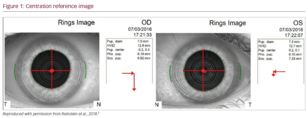
To perform a comprehensive preoperative assessment, it is important to understand the range of imaging technologies available, their nomenclature and their capabilities. Classic ways to screen candidates for refractive surgery include central corneal thickness measurement and Placido disc topography, among others. It is wise to use a combination of imaging techniques, as this will provide a more complete picture of the preoperative landscape than that of a single measure. For example, biomechanical assessment plus tomographic assessment yields a far greater level of detail than simply looking at tomography or front-surface topography data. A more thorough preoperative screening can be a valuable tool when deciding on the most effective refractive procedure.
As mentioned, preoperative forme fruste keratoconus can result in postoperative corneal ectasia; therefore, it is important to define this risk before surgery. However, defining forme fruste prior to refractive surgery can be particularly difficult and require a combination of methods, such as corneal tomography, aberration and biomechanical assessment.12–14 The Ectasia Risk Score System (ERSS) can be used preoperatively to predict the risk of developing ectasia after laser-assisted in-situ keratomileusis (LASIK).6 Preoperative measures include patient age, gender, spherical equivalent refraction, pachymetry and topographic patterns. The ERSS also takes into account perioperative characteristics, including the type of surgery performed, flap thickness, ablation depth and residual stromal bed thickness; and postoperative characteristics, including the time to onset of ectasia. This system has been validated in a retrospective analysis of eyes after LASIK.10 The analysis found that 92% of eyes that developed ectasia were correctly classified as being at high risk, based on the ERSS; however, there was an 8% incidence of false-negatives and 6% incidence of false-positives. Importantly, the ERSS classified significantly more eyes as being a high risk than traditional screening methods (92% versus 50%; p<0.00001).10
More recent methods of screening have also shown higher accuracy to detect mild ectasia with artificial intelligence. The Pentacam Random Forest Index (PRFI) was developed using artificial intelligence to improve detection of ectasia susceptibility based on tomographic data from a multicentre case-controlled study.11 The tomographic and biomechanical index (TBI), a combination of Scheimpflug-based corneal tomography and biomechanics for the detection of preoperative forme fruste, has demonstrated 90.4% sensitivity and 96.0% specificity for subclinical ectasia (optimised TBI cut-off 0.29).13 Additionally, the PRFI method of screening for postoperative ectasia has shown an impressive 100% sensitivity for clinical ectasia.15 While different studies have shown lower accuracy in novel populations of subclinical ectasia cases with normal tomography and normal topography,16,17 a novel optimised artificial intelligence algorithm has been developed to further improve accuracy (unpublished data, to be presented in Paris, France at the 37th Congress of the European Society of Cataract and Refractive Surgeons [ESCRS], September 2019).
Conclusions
The importance of effective preoperative screening for corneal refractive surgery cannot be overstated. Not only can screening assist the surgeon in choosing the safest, most efficient surgical technique, but it can also help predict and plan for any potential complications and provide prognostic information to the patient. It is worth remembering that conditions such as corneal ectasia can also develop as a result of external factors, such as ocular trauma and eye rubbing.11 It is therefore wise to not only assess for predictors of this condition, but also the individual’s susceptibility for progression post-surgery.


