Glaucoma is defined as a chronic, progressive optic neuropathy with loss of retinal ganglion cells and their nerve fibres. One of the most important and the only modifiable risk factor is intraocular pressure (IOP), which gets higher than the (unknown) individual tolerance level (of the optic disc). IOP is not a static value during 24 hours. The fluctuations are of varying amounts in healthy and glaucomatous eyes.1 IOP is subject to a chronobiological pattern and is influenced by changes of head position, emotions, sports activities or the playing of wind instruments.
Glaucoma is defined as a chronic, progressive optic neuropathy with loss of retinal ganglion cells and their nerve fibres. One of the most important and the only modifiable risk factor is intraocular pressure (IOP), which gets higher than the (unknown) individual tolerance level (of the optic disc). IOP is not a static value during 24 hours. The fluctuations are of varying amounts in healthy and glaucomatous eyes.1 IOP is subject to a chronobiological pattern and is influenced by changes of head position, emotions, sports activities or the playing of wind instruments.
The gold standard for measuring IOP is Goldmann applanation tonometry, usually used once during the daytime, which gives information over a period of about five seconds. In patients with progressive glaucoma or in patients with advanced glaucoma a so-called diurnal curve is performed. This means three to five IOP measurements are taken as a minimum within office hours from Monday to Friday. Does this provide enough information about IOP? There is only a 60 % chance to identify peak IOPs of individuals between 08:00 and 16:00.2 Almost nothing is known about IOP during sleeping hours (at least one-third of a day) except a few data from sleep-laboratory studies.3,4 These sophisticated examinations showed that the lowest IOP occurred in the final wake measurement in the light, and the peak IOP occurred in the final measurement in the dark. An increased amount of information is possible by repeated self-tonometry (only when awake), permanent continuous monitoring (inside the eye with an intraocular lens) or temporary continuous monitoring (contact lens).
External Intraocular Pressure Measurement by a Contact Lens
When the distensibility of rabbit eyes was measured using circumference gauges, changes were found with an increase in IOP. Distensibility was slightly higher in the sclera than in the cornea over the range 30–90 mmHg. The change in the angle where the cornea joins the sclera was about 0.020–0.016 radians per mmHg over the range 10–45 mmHg.6 Greene and Gilman were the first to use strain gauges embedded in customised contact lenses in rabbit eyes.7 Strain gauges are made of specific metals (e.g., platinum) or silicon and a strain induces an increase of the resistance (in Ohms) measured by a Wheatstone bridge. The miniaturisation of microchips and progress in radiofrequency transmission have helped technicians to optimise the devices.
Triggerfish
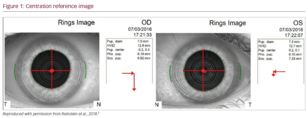
Leonardi et al. developed the idea of a contact lens with embedded strain gauges and called their product Triggerfish (Sensimed, Switzerland).8,9 It is a silicone contact lens with a diameter of 14.4 mm and contains two active strain gauges made out of platinum–titanium (wire loops of 7 μm diameter), one of diameter 11.5 mm, two passive strain gauges for temperature compensation, one antenna (gold, 30 μm) and one microprocessor (50 μm thick) (Figure 1). The contact lens is coated with a hydrophilic substance for better tolerability. Power and data transfer work telemetrically and are wireless from and to an antenna, worn as a ring around the orbit (Figure 2). This antenna is connected to a small recorder, which switches off automatically after 24 hours. The measurements take place in intervals of 8.5 minutes for a period of 1.5 minutes (older software) or in intervals of 5.0 minutes for one of 1.0 minute (new software), which results in 144 or 240 measurements, respectively. The result is a profile in arbitrary units, because the contact lens measures only the change of the curvature of the peripheral cornea caused by changes of the IOP and not the IOP per se. No nomogram exists to convert these measurements. Artefacts caused by blinking or very bright light are filtered.
The tolerability and the safety of the contact lenses were good in patients with ocular hypertension and glaucoma. None of our patients (18 eyes),10,11 but two out of 15 patients in Mansouri and Shaarawy´s series12 discontinued the recording over 24 hours (one patient had severe dry eye, the monitoring of another was interrupted after 17 hours). These authors described ‘significant’ and ‘important’ fluctuations in two of the 24-hour profiles despite different scales on the y-axis and without a definition of a fluctuation – one or more points of measurements? The authors concluded that this technology could possibly lead to improved care of glaucoma patients. Good tolerability and functionality were confirmed also by De Smedt et al.13 These authors examined 10 healthy volunteers to evaluate the tolerability and comfort by a comfort score and the reliability of signal transmission over a 24-hour period. There was no increase in corneal thickness, but there was a low shift to myopia. Immediately after removal of the Triggerfish the visual acuity was reduced. In seven out of the 10 subjects, the Triggerfish was decentred in the vertical axis, which may be the result of a too-tight fit.
Two Other Approaches
The same method of indirect measurement of the IOP by recording changes in the peripheral cornea of pig eyes and of one human volunteer (for two hours) was used by a Spanish group.14 Instead of metallic strain gauges they used a sensor film based on a conducting all-organic nanocomposite bilayer film. The device is a prototype and still wire connected and showed good correlations between changes of resistance and IOP in rapid IOP changes.
The third group, from the University of California, Davis, used polydimethylsiloxane elastomers and modified the material with powdered silver in a predetermined pattern to create conductive wires.15 So far, the patterns are located centrally and therefore disturb vision. No animal or human trials are published.
Usefulness of Results?
There are still no clinical data as to the reproducibility of these results. Leonardi et al.9 carried out their laboratory work to prove the functionality of the device on enucleated porcine eyes; they increased the IOP in rapid shots and obtained a good correlation with the transmitted profile of the contact lens sensor. However, does this reflect reality? Do we have very short and rapid changes in our IOP? Yes, we do, but rarely. We do it in the Valsalva manoeuvre, when playing wind instruments or in yoga exercises with the head upside down. Is this contact lens able to measure changes at the peripheral cornea that result from very slow diurnal, chronobiological changes of the IOP? How much influence does the thickness or the viscosity of the cornea have? There are no data on correlation between corneal hysteresis and Triggerfish profiles. How high is the dependence on a perfect fitting of the contact lens to avoid the lens slipping off the limbal region? We tried to repeat Leonardi´s experiments and used enucleated human eyes instead of porcine eyes. We could not produce a corresponding profile when we increased the IOP of the globe stepwise for 10 minutes. We obtained good correlations only when we increased the IOP rapidly over seconds. In situ examinations differ totally from those of enucleated eyes because of adaptive changes that influence IOP, such as IOP-dependent outflow, ciliary body volume, etc., which are seen with longer term fluctuations but are not encountered in short, rapid changes caused by Valsalva, posture, etc. Additionally, we checked habitual (vertical, horizontal, head down) changes of IOP and obtained no correlations with the IOP.16
In summary, in our opinion, the Triggerfish is not yet ready for everyday use in an ophthalmological office. Several controlled examinations are needed before we can interpret effectively the profile obtained by the wireless contact lens sensor. We have to know what noise there is and what artefacts there are. We also have to examine different qualities of the cornea, too. Safety, tolerability and biocompatibility with the prolonged use seem to be good.
Dynamic Contour Tonometry
As Goldmann tonometry is dependent on the central corneal thickness, dynamic contour tonometry (DCT) has been established successfully. The IOP is measured by a piezoresistive pressure sensor at the centre of the tonometer. It is a membrane that changes its contour according to the pressure difference between both sides and so changes electrical resistance. The same pressure sensor was embedded in the centre of a rigid gas-permeable contact lens (Ziemer Ophthalmic Systems, Switzerland) and measured the IOP 100 times per second.17 A limitation of this device is still a sealed wire connection to the electronic base, which necessarily makes the device unsuitable for IOP measurements over a longer period of time. Additionally, blinking may produce artefacts of the signal. Profiles taken within 100 seconds were reproducible and Valsalva manoeuvres showed perfectly the changes in the IOP. Hedinger et al. stated that an individual radius of the contact lens is mandatory to obtain reliable results.17 Telemetric data transmission is planned. Twa et al. extended the time of measurement with this contact lens to 10 minutes and found comparable data to those of conventional IOP measurements with DCT in sitting subjects.18
Not Yet Suitable for Everyday Use
In summary, IOP-sensing contact lenses are not yet technically mature tools in managing glaucoma. They work on different approaches (strain gauge versus piezoresistance). One measures the changes of the corneal curvature, the other the IOP. So their results are different (changes in resistance expressed by an arbitrary unit versus IOP value). Higher comfort is guaranteed by a wireless transmission of data. Reproducibility of the results is mandatory, but clinical data are missing. However, all ophthalmologists are still curious as to IOP behaviour without therapy, with therapy, after a change of therapy, during night and/or sleep time, during physical activities, in comparison to blood pressure – so we must continue to improve these devices. ■


