Q: What is the optimal procedure for pre- and intraoperative
imaging of the eye during toric IOL implantation?
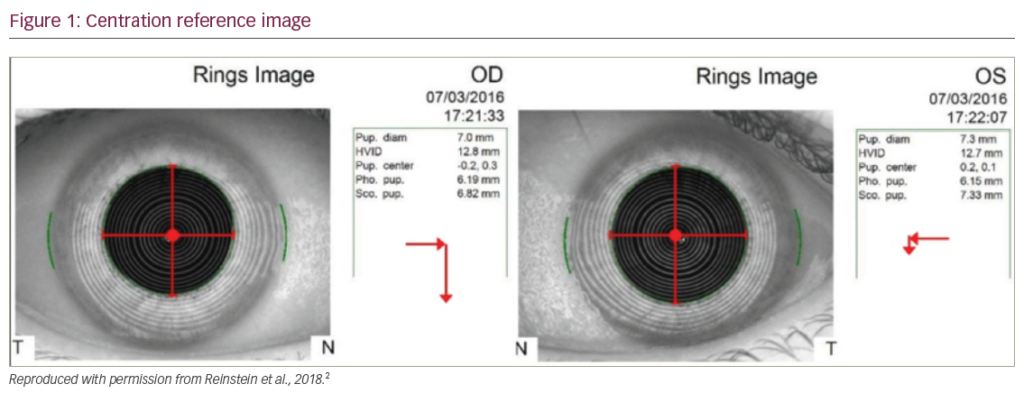
Toric IOLs have perhaps enjoyed the greatest reproducible success of any premium lens type in
use over the past decade. However, their success depends greatly on accurate placement, and this
requires some effort on the part of the surgeon. Even simple measures, such as marking the limbus
pre-operatively with a marking pen, are generally sufficient in order to achieve consistently good
results. However, technology has definitely made an impact in this area. We are currently able to
image the cornea pre-operatively using advanced topography and biometry to take into consideration
the effect on posterior corneal astigmatism on our outcomes. We also have the ability to identify
landmarks on the ocular surface and in some cases project an image into the oculars of the operating
microscope in order to identify the correct axis of implantation.
Q: What is the role of aberrometry during anterior segment imaging?
Ray-tracing aberrometry plays a large role in our clinic in managing various aspects of refractive
surgery planning. We are able to identify the optimal procedure for patients—whether cornea-based
or lens-based, depending on the localization of the aberrations within the eye. In addition, aberrometry
is invaluable in troubleshooting complaints following refractive or cataract surgery especially with
multifocal IOLs. It is also an important patient education tool to help them understand the problems
of their visual system before surgery.
Q: How can the use of corneal imaging techniques improve
outcomes after refractive surgery?
The newest phase of refractive surgery worldwide, and more recently in the US, involves using
topography to plan and execute a refractive treatment that takes into consideration, not only the
lower order aberrations of sphere and cylinder, but also higher order corneal aberrations. Surgeons
internationally have been able to use this technology not only in “normal”
eyes but also in eyes that have had complications from prior keratorefractive
surgery or in cases of corneal ectasia.
Q: In what other ways can OCT be used in cornea
and refractive surgery?
We have been using OCT in several ways. Primarily we are using it as a way
to identify the post-operative anatomy of Descemet membrane endothelial
keratoplasty (DMEK) and Descemet’s stripping automated endothelial
keratoplasty (DSAEK) graft adherence. We have also been using the OCT
to calculate the true corneal power, and this has made a huge impact in
our post-refractive surgery IOL calculations. A newly available modality
will be epithelial thickness mapping. Although available for many years
internationally, surgeons in the US have only recently had widespread
access to this.
Q: How important is the use of intraoperative OCT?
Intraoperative OCT may be of benefit in DMEK and DSAEK cases as
a way to help identify correct orientation of the tissue as well as ideal
positioning. There may also be uses in posterior segment (retina) surgery.
At the moment, while it is a nice tool, it is cost-prohibitive for the majority
of surgeons.


